- MAGNETOM World
- Hot Topics
- Lower-field MRI
Lower-field MRI
For more than 20 years, the clinical standard for MR imaging has been either 1.5T or 3T. While many of the physical advantages of low field have been well known in the scientific community, the push for higher SNR favored higher fields. However, in the light of technical improvements, other metrics may be favored. Improvements in image reconstruction – from parallel imaging and compressed sensing to deep learning – mean that clinicians can make optimal use of the available signal while exploiting the physical advantages of low-field MRI, such as reduced artifacts.
If SNR is sufficient at two different fields, then the focus may fall instead on diagnostic or financial value.
Scientific literature
even back in the mid 1990s showed no significant difference in diagnostic
sensitivity or specificity between 1.5T and lower-field systems1.
As scanner costs typical scales with field strength,
lower field scanners offer opportunities given the cost pressure in many
healthcare systems. All these factors could
help bring MRI to places it has not been before – spreading into new geographical and clinical areas.
Clinical Experience

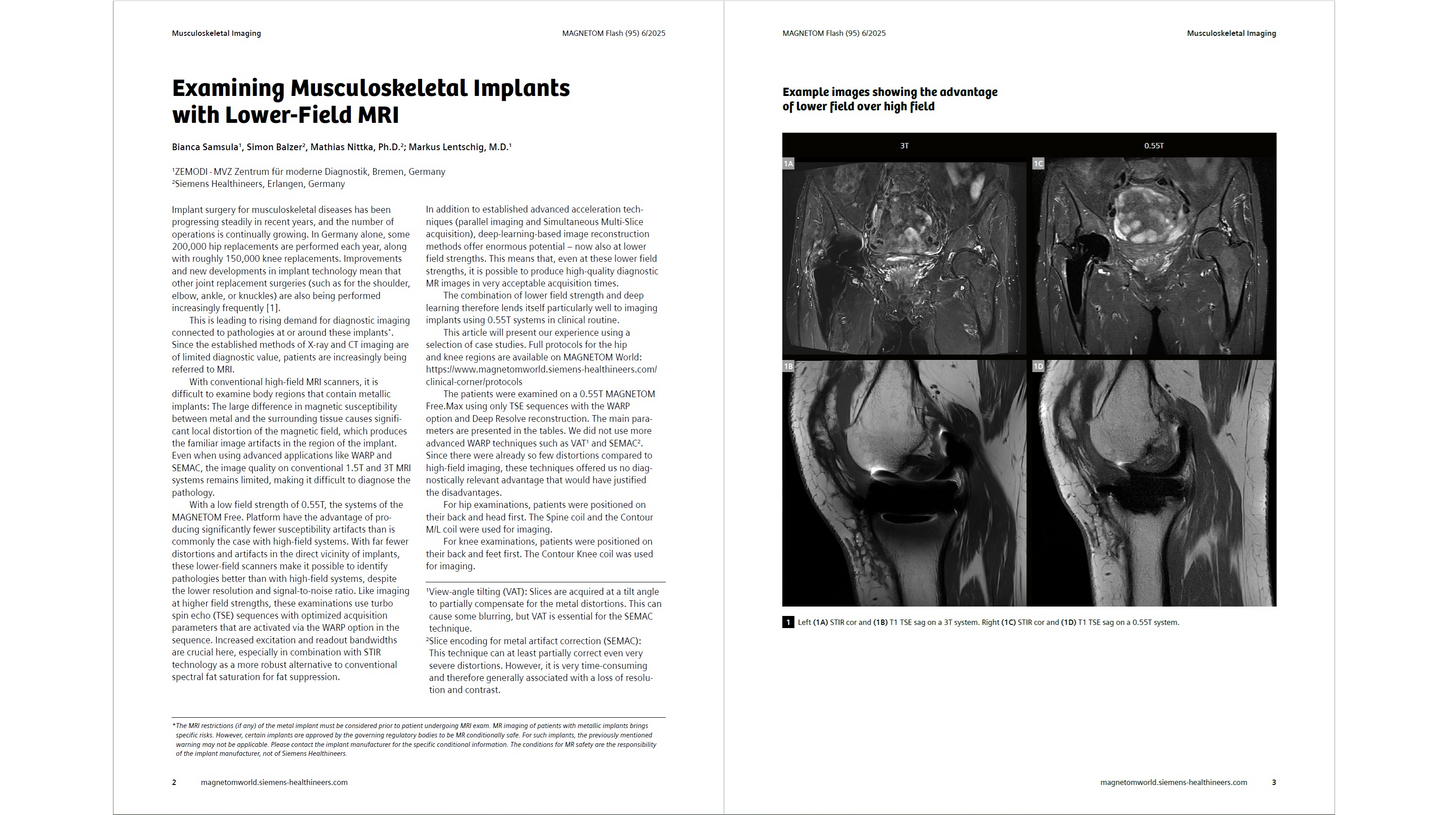
Examining Musculoskeletal Implants with Lower-Field MRI
Bianca Samsula; Markus Lentschig, M.D.; et al.
ZEMODI - MVZ Zentrum für moderne Diagnostik, Bremen, Germany

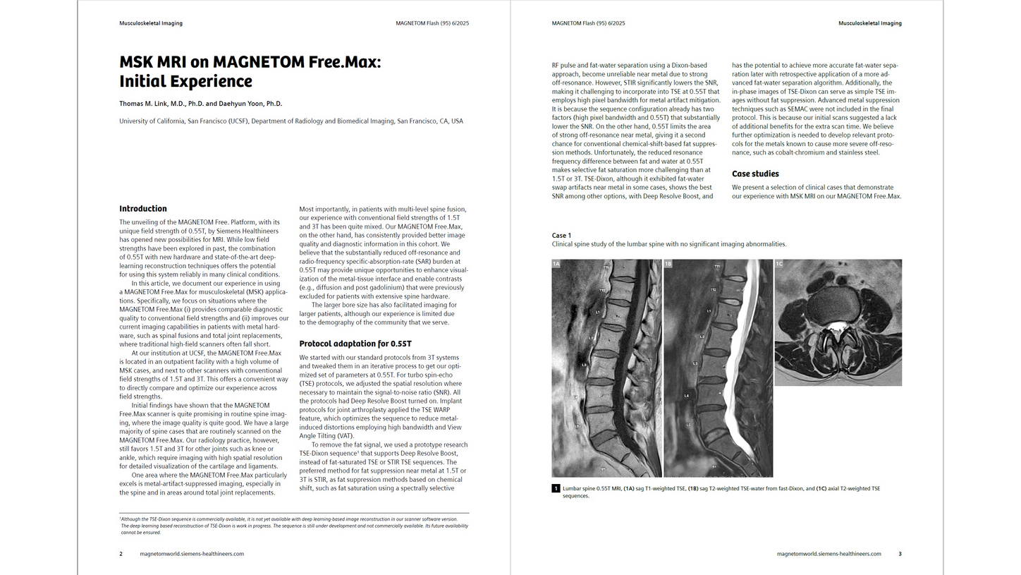
MSK MRI on MAGNETOM Free.Max: Initial Experience
Thomas M. Link, M.D., Ph.D. and Daehyun Yoon, Ph.D. University of California, San Francisco (UCSF), Department of Radiology and Biomedical Imaging, San Francisco, CA, USA

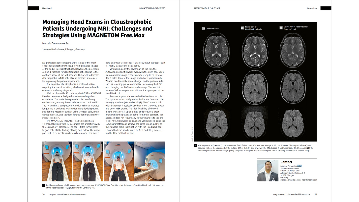
Managing Head Exams in Claustrophobic Patients Undergoing MRI: Challenges and Strategies Using MAGNETOM Free.Max
Marcelo Fernandes Arêas
Siemens Healthineers, Erlangen, Germany

Establishing an MRI-Guided Intervention Service Using a 0.55T System: Initial Experiences and Workflow Development
Jan Robert Kroeger, M.D.; et al. (Johannes Wesling Klinikum Minden, University Hospital of Ruhr University Bochum, Germany)

Clinical Implementation of 0.55T MRI Simulation for Stereotactic Radiotherapy Using the MAGNETOM Free.Max RT Edition
Joshua Kim, Ph.D.; Kundan Thind, Ph.D.; et al. (Department of Radiation Oncology, Henry Ford Health, Detroit, MI, USA)

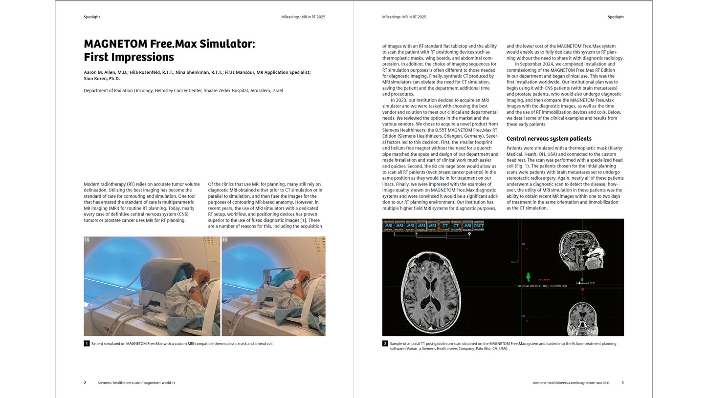
MAGNETOM Free.Max Simulator: First Impressions
Aaron M. Allen, M.D.; et al. (Department of Radiation Oncology, Helmsley Cancer Center, Shaare Zedek Hospital, Jerusalem, Israel)

Experience with 0.55T MRI: Case Series Highlighting Claustrophobic, Metal Implant, Pediatric, and Follow-Up Scenarios
Ercan Karaarslan, M.D.; et al. (Acıbadem Mehmet Ali Aydınlar University, Faculty of Medicine, İstanbul, Turkey)

Susceptibility-Weighted Imaging at Lower Field Strength with Deep Learning Image Reconstruction
Johanna Maria Lieb, M.D.; et al. (University Hospital Basel, Switzerland)

Stroke Case Study Using a Lower-Field MRI System in a Tier 4 Town of Southern India
RG Jyothi, DNB; et al. (Jyothi Diagnostics, Madanapalle, India)

MAGNETOM Free. Platform special issue

Imaging Titanium and Carbon Fiber Spinal Implants at 0.55T, 1.5T, and 3T: A Phantom Experiment
Xin Miao, et al. (Siemens Healthineers, Boston, MA, USA)
Exploring the Potential of 0.55T MRI: Triumphs, Trials, and Future Horizons
Michael Bach, M.D. (University Hospital Basel, Switzerland)
Siemens Healthineers Lunch Symposium, ESMRMB 2023, Basel, Switzerland

Brachytherapy Treatment Planning for Cervical Cancer Patients Using a Lower Magnetic Field MR Scanner

Low Field MRI Impact on Interventions
Paul J.A. Borm, Ph.D.; et al. (Heinrich Heine University (HHU), Düsseldorf & Nano4Imaging GmbH, Aachen, Germany)

MAGNETOM Free.Max. The Power of Deep Resolve
Heike Weh and Thomas Illigen (Siemens Healthineers, Erlangen, Germany)

Intrahepatic Cholangiocarcinoma: Typical Imaging Findings at 0.55T

MAGNETOM Free.Star: Initial Experience in a Tier 3 Indian City
V. Suresh, M.D. (Dolphin Diagnostic Centre, Vishakhapatnam, India)

Fetal Low Field MRI – the First 150 Cases
Jana Hutter, Ph.D.; et al. (Centre for the Developing Brain, School of Biomedical Engineering, King’s College London, UK)

Exploring the Potential of Low-Field Musculoskeletal MRI at 0.55T: Preliminary Results in Patients with Large Metal Implants
Hanns-Christian Breit, M.D.; et al. (Dept. of Radiology, University Hospital Basel, University of Basel, Switzerland)

Cardiac MRI on the MAGNETOM Free.Max: The Ohio State Experience
Orlando P. Simonetti, PhD, MSCMR, FISMRM, FAHA; et al. (The Ohio State University, Columbus, OH, USA)

Improving the Assessment of the Postoperative Spine with 0.55T MRI: A Case Report
Hanns-Christian Breit, M.D.; et al. (Dept. of Radiology, University Hospital Basel, University of Basel, Switzerland)
Opportunities at 0.55 Tesla
Krishna S. Nayak, Ph.D. (University of Southern California, Los Angeles, CA, USA)
ISMRM Lunch Symposium 2021

Image Gallery 0.55T MAGNETOM Free.Max – Breaking Barriers

Experience of Using a New Autopilot Assistance System for Easy Scanning in Brain and Knee MRI Examinations
Tanja Dütting, Stephan Clasen
(Institute for Diagnostic and Interventional Radiology, Kreiskliniken Reutlingen, Germany)

Iterative Denoising Applied to 3D SPACE CAIPIRINHA:
A New Approach to Accelerate 3D Brain Examination in Clinical Routine
A New Approach to Accelerate 3D Brain Examination in Clinical Routine
Alexis Vaussy, Thomas Troalen, et al.
(Siemens Healthineers, Saint-Denis, France)

The Next Generation – Advanced Design Low-field MR Systems
Val M. Runge and Johannes T. Heverhagen
(University Hospital of Bern, Inselspital, University of Bern, Switzerland)

Magnetic Resonance Fingerprinting for Precision Imaging in Neuro-Oncology
Domenico Zacà; Guido Buonincontri
(Siemens Healthineers, Italy)
Low-, Mid-, and High-Field MRI.
A Critical Historical Review and How Challenges at Lower-Field Strength MRI can be Tackled with Emerging Technologies
(University Hospital Bern, Inselspital, Switzerland)
(Re-)Emerging Fields of Lower-field MRI
Hersh Chandarana (NYU Langone Health, New York, NY, USA)

Brain MRI in an Emergency Department:
Clinical Implementation and Experience in the First Year
Clinical Implementation and Experience in the First Year
Vincent Dunet, et al.
(Lausanne University Hospital and University of Lausanne, Switzerland)
Bringing MRI to the People. What about the ICU?
Elmar Merkle (University Hospital Basel, Switzerland)

GOBrain in Acute Neurological Emergencies:
Diagnostic Accuracy and Impact on Patient Management
Philipp M. Kazmierczak, et al.
(Klinik und Poliklinik für Radiologie, Klinikum der Universität München, Germany)

Cardiopulmonary Imaging Using a High-Performance 0.55T MRI System
Adrienne E. Campbell-Washburn; Robert J Lederman; Robert S. Balaban
(Division of Intramural Research, National Heart, Lung, and Blood Institute, National Institutes of Health, Bethesda, MD, USA)
MRI-guided Cardiovascular Catheterization at 0.55T
Adrienne Campbell-Washburn
(NIH, Nat. Heart, Lung, and Blood Institute, Burnsville, MN, USA)
Opportunities in Interventional and Diagnostic Imaging by Using High-performance Low-field-strength MRI
Adrienne Campbell-Washburn (NIH, Nat. Heart, Lung, and Blood Institute, Bethesda, MD, USA)
Lung MRI of COVID-19 Patients
Sebastian Bickelhaupt (University Hospital Erlangen, Germany)
Technology

Real-Life Considerations and Benefits of the AutoResponse Feature in MRI Systems with DryCool Technology
Yatin Sharma, et al. (Siemens Healthcare Pvt Ltd, India)

Our Connected Scanner is a Smart Deal: You Care for Your Patients, We Stay Connected to Care for Your MAGNETOM Free.Max
Julia Sauernheimer (Service Business Manager for MRI, Siemens Healthineers, Erlangen, Germany)

MAGNETOM Free.Max: Keeping a Hot System Cool
Stephan Biber, Ph.D. (Senior System Architect at Siemens Healthineers, Erlangen, Germany)

MAGNETOM Free.Max: How to Make it Big Inside & Small Outside
Stephan Biber, Ph.D. (Senior System Architect, Siemens Healthineers, Erlangen, Germany)

MAGNETOM Free.Max: from Concept to Product, a Brief History of the DryCool Magnet Development
Simon Calvert, CEng FIMechE (Head of Product Innovation & Chief Technology Officer, Siemens Healthineers, Oxford, UK)

Revisiting the Physics behind MRI and the Opportunities that Lower Field Strengths Offer
André Fischer, Ph.D.
(Siemens Healthineers, Erlangen, Germany)
Access to MRI - for everyone, everywhere

Gradient-Free MRI for the Moon and Other Hard-To-Reach Places
Gordon E. Sarty, Ph.D.; et al. (Space MRI Lab, QuanTA Centre, University of Saskatchewan, Saskatoon, Canada)

Building a Global Power of Experience in Diagnostic Imaging - Lessons from Africa's COVID-19 Response
Udanna Anazodo, Ph.D.; et al. (Montreal Neurological Insitute, McGill University, Montreal, Quebec, Canada)
Accessible MRI - the Clinical View
Vikas Gulani, M.D., Ph.D. (University of Michigan, Ann Arbor, MI, USA)
Quantitative and Intelligent MRI for Underserved Populations
Nicole Seiberlich, Ph.D. (University of Michigan, Ann Arbor, MI, USA)

Current Status of MRI in India
Raju Sharma, MD; Devasenathipathy Kandasamy, MD; and Ankur Goyal, MD (All India Institute of Medical Sciences, New Delhi, India)
Accessible MRI - Technological and Economic Challenges
Najat Salameh, Ph.D. and Mathieu Sarracanie, Ph.D. (Center for Adaptable MRI Technology, University of Basel, Switzerland)

Re-Envisioning Low-Field MRI
Najat Salameh, Ph.D. and Mathieu Sarracanie, Ph.D.
(Center for Adaptable MRI Technology, Department of Biomedical Engineering, University of Basel, Switzerland)

MAGNETOM Free. Platform special issue

Download the MAGNETOM Flash special issue on MAGNETOM Free.Max
Did this information help you?
Thank you.
Lee DH et al. MR Imaging Field Strength: Prospective Evaluation of the Diagnostic Accuracy of MR for Diagnosis of Multiple Sclerosis at 0.5 and 1.5T. Radiology. 1995;194:257-262.
Vellet AH et al. Anterior cruciate ligament tear: prospective evaluation of diagnostic accuracy of middle- and high-field-strength MR imaging at 1.5 and 0.5 T. Radiology. 1995;197:826-830.